Currently, ulcerative sexually transmitted infections, including syphilis, herpes simplex virus (HSV), lymphogranuloma venereum (LGV), chancroid, donovanosis and, more recently, monkeypox (MPOX), represent a growing challenge for health care professionals.
The incidence of syphilis and LGV has increased in recent years in Spain. Additionally, HSV, syphilis and chancroid can also increase the risk of HIV acquisition and transmission. The population groups most vulnerable to these infections are young people, men who have sex with men (MSM) and commercial sex workers.
It is important to make a timely differential diagnosis since genital, anal, perianal, and oral ulcerative lesions may pose differential diagnosis with other infectious and non-infectious conditions such as candidiasis vulvovaginitis, traumatic lesions, carcinoma, aphthous ulcers, Behçet's disease, fixed drug eruption, or psoriasis. For this reason, the dermatologist plays a crucial role in the diagnosis and management of sexually transmitted infections.
This chapter presents the main epidemiological, clinical and therapeutic features associated with these infections.
En la actualidad, las infecciones de transmisión sexual ulcerativas, que incluyen la sífilis, el virus del herpes simple (VHS), el linfogranuloma venéreo (LGV), el chancroide, la donovanosis y, más recientemente, el monkeypox (MPOX), representan un desafío creciente para los profesionales sanitarios.
La incidencia de sífilis y LGV ha aumentado en los últimos años en España. Además VHS, sífilis y chancroide son infecciones que pueden incrementar el riesgo de la adquisición y transmisión de VIH. Los grupos poblacionales con mayor vulnerabilidad ante estas infecciones son jóvenes, hombres que tienen sexo con hombres (HSH) y trabajadores del sexo comercial.
Es importante realizar un diagnóstico diferencial oportuno, ya que las lesiones ulcerativas genitales, anales, perianales y orales pueden plantear diagnóstico diferencial con otras condiciones infecciosas y no infecciosas, como vulvovaginitis candidiásica, lesiones traumáticas, carcinoma, aftas, enfermedad de Behçet, eritema fijo medicamentoso o psoriasis. Por este motivo, el dermatólogo tiene un papel crucial en el diagnóstico y el tratamiento de las infecciones de transmisión sexual.
En el presente capítulo se exponen las principales características epidemiológicas, clínicas y terapéuticas de dichas infecciones.
The development of these guidelines is justified by the increasing rise in sexually transmitted infections (STIs), particularly among adolescents, men who have sex with men (MSM), and commercial sex workers.
It is important to establish an appropriate differential diagnosis in each case, apply the relevant microbiological techniques based on sexual exposure and administer effective treatment for each of them.1
Syphilis/LuesGiven its clinical and epidemiological relevance, the etiopathogenesis and clinical signs of syphilis will be addressed in the chapter “Guidelines on the management of syphilis” (which will be available in upcoming publications of this journal), including in this section only its diagnosis and treatment.
DiagnosisThe definitive method for diagnosing both primary and congenital syphilis is the identification of Treponema pallidum via dark field microscopy or through the application of molecular tests (PCR) on an exudative lesion or tissue. However, these techniques are not universally distributed and are only available in specific centers, so the diagnosis of syphilis at any stage continues to be based on treponemal and non-treponemal serological tests.2
Treponemal tests (T. pallidum passive particle agglutination [TP-PA] assay or FTA-ABS, etc.) are the most specific and the first to test positive; however, once they have tested positive, they do not go back to negative (in most cases), so their fluctuations are not useful for follow-up or new diagnoses.3 Since they are more sensitive to non-treponemal tests (Venereal Disease Research Laboratory [VDRL] or Rapid Plasma Reagin [RPR] test) false positives may occur more frequently, and they may take a few more days to test positive than treponemal tests. However, with appropriate treatment, they can go back to negative, so their fluctuations and new positivities are useful for the follow-up and diagnosis of new infections.4
To achieve a correct diagnosis, it is necessary to perform both tests.
To say that an infection is properly treated, a decrease of at least 2 dilutions (e.g., 1:16 down to 1:4, 1:32 down to 1:8) is required 6 months into therapy. The same test should be used for both diagnosis and follow-up, and if possible, in the same laboratory3 (2, C).
If both tests are positive, treatment is prescribed based on the patient's clinical stage. If the treponemal test is positive and the non-treponemal one is negative, it is advisable to perform another treponemal test using a different method; if it remains positive and there is a previous history of syphilis, no further study is recommended. If there is no prior history and/or the clinical or epidemiological suspicion is high, it is recommended to retest in 2–4 weeks, emphasizing the importance of sexual abstinence until the case is resolved.2
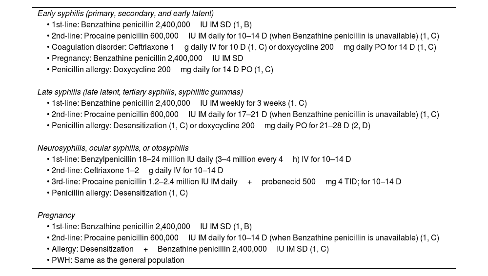
TreatmentThe treatment of syphilis is based on staging (Table 1). In all cases, the antibiotic of choice is parenteral penicillin, whose efficacy is supported by extensive clinical experience and clinical trials. There are some situations in which the administration of another antibiotic is permitted, with stricter follow-up requirements.
Syphilis treatment by stage and evidence level.
| Early syphilis (primary, secondary, and early latent) |
| • 1st-line: Benzathine penicillin 2,400,000IU IM SD (1, B) |
| • 2nd-line: Procaine penicillin 600,000IU IM daily for 10–14 D (when Benzathine penicillin is unavailable) (1, C) |
| • Coagulation disorder: Ceftriaxone 1g daily IV for 10 D (1, C) or doxycycline 200mg daily PO for 14 D (1, C) |
| • Pregnancy: Benzathine penicillin 2,400,000IU IM SD |
| • Penicillin allergy: Doxycycline 200mg daily for 14 D PO (1, C) |
| Late syphilis (late latent, tertiary syphilis, syphilitic gummas) |
| • 1st-line: Benzathine penicillin 2,400,000IU IM weekly for 3 weeks (1, C) |
| • 2nd-line: Procaine penicillin 600,000IU IM daily for 17–21 D (when Benzathine penicillin is unavailable) (1, C) |
| • Penicillin allergy: Desensitization (1, C) or doxycycline 200mg daily PO for 21–28 D (2, D) |
| Neurosyphilis, ocular syphilis, or otosyphilis |
| • 1st-line: Benzylpenicillin 18–24 million IU daily (3–4 million every 4h) IV for 10–14 D |
| • 2nd-line: Ceftriaxone 1–2g daily IV for 10–14 D |
| • 3rd-line: Procaine penicillin 1.2–2.4 million IU IM daily+probenecid 500mg 4 TID; for 10–14 D |
| • Penicillin allergy: Desensitization (1, C) |
| Pregnancy |
| • 1st-line: Benzathine penicillin 2,400,000IU IM SD (1, B) |
| • 2nd-line: Procaine penicillin 600,000IU IM daily for 10–14 D (when Benzathine penicillin is unavailable) (1, C) |
| • Allergy: Desensitization+Benzathine penicillin 2,400,000IU IM SD (1, C) |
| • PWH: Same as the general population |
D: day(s); SD: single dose; IM: intramuscular; IV: intravenous; PO: oral route; TID: times per day.
The GRADE methodology was used for the formulation of the recommendations.
- •
Contact with a patient with early syphilis<90 days from contact: treat as early syphilis, even if serological tests are negative.
- •
Contact with a patient with early syphilis>90 days from contact: if tests are negative, do not treat; if tests are positive, treat based on staging; if dealing with a patient at risk of being lost to follow-up, treat as early syphilis.
- •
Contact with a patient with late syphilis: if tests are positive, treat based on their particular staging (Table 2).
Table 2.Syphilis follow-up by stage and evidence level.
Early syphilis: VDRL/RPR testing at 3, 6, and 12 months, with a minimum decrease of 2 dilutions (4-fold). If no decrease, consider the possibility of neurosyphilis (2, D). Late syphilis: VDRL/RPR may not test negative. Follow-up is not recommended unless there is a high risk of reinfection.
Herpes infection is characterized by the fact that, after the primary infection, the virus remains latent and can reactivate throughout life.2 Both herpes simplex virus type 1 (HSV1) and 2a (HSV2) can affect the genital area, with HSV2 being the most common one. Many patients remain undiagnosed due to low suspicion in the case of pauci or asymptomatic herpes, and these patients are at high risk of transmitting the infection.
The risk of transmission appears to be higher during prodromal symptoms and recurrences, so patients should be advised to abstain from sexual contact during these periods. The efficacy of condoms in preventing transmission has not been proven; however, indirect data support their use in both women and men5 (IIb, B).
Clinical presentationClinically, it is characterized by painful, recurrent, and self-resolving vesicles and ulcers. Confirmatory diagnosis with PCR is advised whenever possible, bearing in mind that most patients seek consultation when the outbreak has already resolved6 (IIIb). Recurrences and subclinical forms in the genital area are more frequently described with HSV2 vs HSV1.7
DiagnosisThe recommended diagnostic test is PCR due to its high sensitivity5 (Ib, A); given the low sensitivity of culture, it is only advised as a diagnostic technique when PCR is not available or for studying antiviral resistance in refractory cases. Samples should be drawn from the active lesion8 (Ib, A); sampling from random areas is ill-advised due to its low sensitivity, and because a negative result does not rule out the possibility of latent infection (Ib, A).
Regarding HSV serologies, their use is recommended only in the following scenarios5:
- 1.
Patients with recurrent lesions or atypical presentation and where PCR or culture tests repeatedly negative or could not be performed (III, B).
- 2.
To differentiate between a first episode or a recurrence for the purpose of counseling and management (III, B).
- 3.
Partners of patients diagnosed with HSV who do not know if they have previously had lesions, for counseling purposes (Ib, A).
- 4.
Asymptomatic pregnant individuals with a partner with a past medical history of herpes (IIb, B).
The goal of treatment in primary infection is to alleviate symptoms and prevent or reduce the frequency of outbreaks; and in recurrences, to reduce the likelihood of transmission. Treatment cannot eradicate the virus, and it has not been shown to affect the frequency or severity of subsequent outbreaks.2
Treatment in primary infectionTreatment is recommended for all patients diagnosed with a primary herpetic infection within the first 5 days of infection or while they have active lesions,5 due to the symptomatic nature of the condition and because it may follow a latent course (Table 3).
Treatment of primary HSV infection.
| Primary HSV infection treatment (Ib, A) |
| • Acyclovir: 400mg PO, 3 TID for 7–10 D (preferred in pregnant women) |
| • Famciclovir: 250mg PO, 3 TID for 7–10 D |
| • Valacyclovir: 1g PO, 2 TID for 7–10 D |
| • Acyclovir: 200mg PO, 5 TID for 7–10 D (not recommended due to poor adherence) |
D: day(s); PO: oral route; TID: times per day.
Topical treatments are not recommended as they are less effective than oral treatments and may lead to resistance9,10 (IV, C). IV treatment is reserved only for patients unable to take oral drugs due to functional disorders or vomiting, and for immunosuppressed patients.
Treatment of recurrencesRecurrences of HSV1 are generally less frequent than HSV2, which highlights the importance of genotyping for post-infection counseling (Table 4).
Treatment of HSV-2 recurrences.
| HSV-2 recurrence treatment (Ib, A) |
| • Acyclovir: 400mg PO, 3 TID for 7 days (not recommended) |
| • Acyclovir: 800mg PO, 2 TID for 5 days |
| • Acyclovir: 800mg PO, 3 TID for 2 days |
| • Famciclovir: 1g PO, 2 TID for 1 day |
| • Famciclovir: 500mg SD+250mg 2 TID for 2 days |
| • Famciclovir: 125mg PO, 2 TID for 5 days |
| • Valacyclovir: 500mg PO, 2 TID for 3 days |
| • Valacyclovir: 1g PO once daily for 5 days |
D: day(s); SD: single dose; TID: times per day; PO: oral route.
The ideal time to start treatment is during the prodrome of the infection or on day 1 of lesion appearance. Recurrence treatment shortens the outbreak duration by 1–2 days5 (Ib, A).
Suppressive treatmentThis should be agreed upon with the patient and is recommended in cases where HSV2 or HSV1 infection is confirmed, and frequent outbreaks occur that impact the patient's quality of life. Suppressive treatment can reduce the possibility of recurrence by 70% up to 80%.5 Additionally, it reduces asymptomatic viral shedding.2 Long-term safety and efficacy have been demonstrated.2 Suppressive therapy is recommended for symptomatic serodiscordant couples or those with multiple partners.5 There is no data on effectiveness in asymptomatic HSV2 patients (Table 5).
Suppressive treatment of HSV-2.
| Suppressive HSV treatment (Ib, A) |
| • Acyclovir: 400mg PO, 2 TID |
| • Valacyclovir: 500mg PO, once daily (not recommended if >10 outbreaks per year) |
| • Valacyclovir: 1g PO, daily (preferred if >10 outbreaks per year) |
| • Famciclovir: 250mg PO, 2 TID |
TID: times per day; PO: oral route.
HSV management in immunosuppressed patients and people living with HIV (PWH) (Table 6). Other special situations are considered in Table 7.
Special considerations, primary HSV infection treatment in PWH.
| Primary HSV infection treatment in PWH (2) |
| • Acyclovir: 400mg PO, 5 TID for 7–10 days (IV, C) |
| • Valacyclovir: 500–1000mg PO, 2 TID for 10 days (IV, C) |
| • Famciclovir: 250–500mg PO, 3 TID for 10 days (IV, C) |
| • Severe disease: acyclovir 5–10mg/kg every 8h for 2–7 days or until clinical improvement, followed by oral antivirals for a minimum of 10 days (IV, C) |
TID: times per day; PO: oral route.
Other special considerations.
| Treatment for suppression in PWH patient (Ib, A) | Treatment for recurrences in PWH patient (Ib, A) | Suppressive treatment for pregnant women (10) (start at week 36) (Ib, A) | Treatment for recurrent herpes in immunocompromised patient (Ib, A) |
|---|---|---|---|
| Acyclovir 400–800mg PO 3 TID | Acyclovir 400mg PO 3 TID 5–10 D | Acyclovir 400mg PO 3 TID (Ib, B) | Accessible lesions: Foscarnet cream or Cidofovir 1% gel |
| Famciclovir 500mg PO 2 TID | Famciclovir 500mg PO 2 TID 5–10 D | Valaciclovir 500mg PO 2 TID | Inaccessible lesions: Foscarnet 40mg/kg IV every 8h or Cidofovir 5mg/kg IV weekly |
| Valaciclovir 500mg PO 2 TID | Valaciclovir 1g PO 2 TID 5–10 D |
D: day(s); IV: intravenous; TID: times per day; PO: oral route.
The most important risk factor for HSV reactivation is the degree of immunosuppression. Systemic antiviral treatment regimens have been shown to be just as effective in immunosuppressed patients; however, antiviral resistance is much more common in PWH, which can lead to more frequent therapeutic failures.5
Lymphogranuloma venereumLymphogranuloma venereum (LGV) is a sexually transmitted infection caused by the bacteria Chlamydia trachomatis serotypes L1–L3, with L2 and L2b being the most common in Europe.11,12 Since 2003, it has been endemic in Europe among men who have sex with men (MSM),13 especially in PWH14 (1,A), and since 2015, it has been classified as a notifiable disease in Spain.
Clinical presentationApproximately 25% of LGV infections in MSM are asymptomatic.15 In symptomatic individuals, the incubation period may vary between 1 and 4 weeks, with 3 classic stages described:
- •
First stage: A small, painless papule or pustule that may ulcerate and resolves on its own within a week. Mucopurulent discharge may occur, especially with rectal involvement.
- •
Second stage (“inguinal stage”): Painful inguinofemoral lymphadenopathy appears 2–6 weeks after the primary lesion, typically unilateral (2 out of 3 cases). Complications may include inflammation, suppuration, and abscesses; some of these may drain spontaneously. The “groove sign” (pathognomonic) occurs due to the inflammation of inguinal and femoral lymph nodes separated by the inguinal ligament.
- •
Third stage (“ano-genito-rectal stage”): Initially presents as proctocolitis, characterized by severe symptoms of anorectal pain, hemopurulent discharge, and rectal bleeding, along with tenesmus and constipation. This is followed by perirectal abscesses, fistulas, or rectal stenosis. It can mimic chronic inflammatory bowel diseases, such as Crohn's disease, both clinically and histopathologically, which should be considered in differential diagnosis. Without treatment, it can lead to complications such as megacolon, chronic progressive lymphangitis with elephantiasis, esthiomene (chronic ulcerative vulvar disease), or frozen pelvis syndrome. Currently, the most common presentation of LGV is symptomatic proctitis in MSM, with genital ulcers or inflammatory inguinal adenopathies being less common.
Less frequently, it may be accompanied by systemic symptoms, such as mild fever, chills, malaise, arthralgia–myalgia, reactive arthritis, pneumonitis, and perihepatitis.
DiagnosisThe initial diagnosis is based on clinical suspicion but should be confirmed by the detection of specific DNA of the LGV variant (L1, L2, and L3), through a 2-step procedure (1, B).16–18
- •
Nucleic acid amplification tests (NAAT) are used on suspected clinical samples.
- •
If C. trachomatis DNA/RNA is detected, LGV-specific genotype DNA should be identified.
Early treatment initiation is recommended to reduce potential complications, such as megacolon and chronic progressive lymphangitis (2,C) (Table 8).
Treatment of lymphogranuloma Venereum.
| 1st choice treatment |
| • Doxycycline:Symptomatic: 100mg every 12h for 21 days PO (1, B)19Asymptomatic: 100mg every 12h for 21 days PO (1, C) or short coursea: 100mg every 12h for 7 days PO (2, D)20 |
| Alternativetreatmentb |
| • Azithromycin: 1g single dose or 1g weekly for 3 weeks PO (2, D)21 |
| • Erythromycin (preferred in pregnant women): 400mg every 6h for 21 days PO (2, D) |
| Surgical treatment |
| • Fluctuating buboes should be drained via needle aspiration (2, D). Surgical incision is not recommended due to risk of complications such as chronic cavity formation (2, D). |
D: day(s); PO: oral route.
In some cases, longer treatment may be required if there are complications such as fistulas or buboes.22
Chancroid, or soft chancre, is a sexually transmitted infection caused by the Gram-negative bacterium Haemophilus ducreyi.23 It is more common in tropical countries, although its incidence has dropped even in most countries where it was previously endemic, except in northern India and Malawi.24,25
Clinical presentationAfter an incubation period of 3–7 days, small, painful erythematous papules appear, which quickly progress to pustules and ulcers. These are characterized by being superficial with irregular, undermined edges, a granulomatous base, and purulent exudate, which can persist for months if untreated. Up to 50% of cases are accompanied by painful unilateral inguinal lymphadenopathy that can coalesce into buboes.
DiagnosisDiagnosis is usually clinical (Table 9), as bacterial culture isolation is difficult (sensitivity<80%) (III,B).26 Nucleic acid amplification techniques, such as PCR, are the most commonly used and can differentiate between different microbiological agents, such as T. pallidum and HSV27,28 (III,B). Treatment details are provided in Table 10.
Probable diagnosis of chancroid (according to CDC).18
| If the following criteria are met |
| 1. The patient has one or more painful genital ulcers. |
| 2. Clinical presentation of genital ulcers and, if present, regional lymphadenopathy is typical of chancroid. |
| 3. No evidence of T. pallidum infection by dark field examination of ulcer exudate or serological testing for syphilis at least 7 days after ulcer onset. |
| 4. PCR testing for HSV-1 or 2 or culture of HSV from ulcer exudate tests negative. |
Patients should be reevaluated 3–7 days after treatment. The healing time depends on the size of the ulcer and lymphadenopathy, and progression is slower in PWH and uncircumcised individuals.29
Contact tracing, including sexual partners from the 10 days preceding symptom onset is advised.
MPOXMonkeypox (MPOX) is an emerging zoonotic disease caused by an orthopoxvirus (double-stranded DNA virus; genus Orthopoxvirus, family Poxviridae).30
It is considered endemic in Central and West Africa, where 2 variants have been identified: the Central Africa/Congo Basin variant (mortality rate around 10%) and the West Africa variant (mortality rate of 3% up to 4%).31–33
In May 2022, several cases of MPOX were identified without recent travel history to endemic areas or contact with confirmed cases, prompting the WHO, on July 23rd of 2022, to declare this latest outbreak as an international public health emergency.34 As of the writing of this article, a total of 91,123 cases had been reported worldwide, with 157 deaths.35 In Europe, 26,229 cases were reported,36 with Spain being the country with the most reported cases (7611).35,36
In endemic areas, there are four forms of transmission of MPOX: bites or scratches from infected animals (squirrels, rodents, monkeys), contact with their fluids, consumption of insufficiently cooked meat, or infected humans. In the latter case, infection can be acquired through the airborne route (microdroplets), direct contact (wounds, scabs, bodily fluids), and indirect contact (fomites), although vertical and nosocomial transmission have also been described.37
In the current outbreak, it is considered that the main mechanism of transmission is sexual contact,38 with the most affected individuals being MSM, young people (mean age of 34 years), and PWH (52.5%).35,36,38–42
Clinical presentationThe incubation period goes from 5 up to 21 days.43 The most common symptom is fever.35,36,38–42 Clinical signs are related to the transmission route44:
- -
Respiratory/Classical (mainly endemic form): initially fever, headache, lymphadenopathy, myalgia; later, multiple papules and pseudopustules.
- -
Subcutaneous inoculation: well-circumscribed, deep pseudopustules, often umbilicated, painful, taking weeks to resolve, which may be accompanied by local edema and regional lymphadenopathy. Lesions may also exhibit perioral/lingual involvement or present as paronychia.
Complications have been reported in up to 8.3% of cases,38–42 with the most common being bacterial superinfection, oral ulcers, and proctitis/proctocolitis/proctalgia.45–51
DiagnosisDiagnosis is clinical–epidemiological and confirmed by PCR and/or sequencing.52
Histopathological analysis may reveal acidophilic cytoplasmic inclusion bodies (“Guarnieri bodies”), which correspond to aggregates of viral particles.
Differential diagnosis should include varicella, herpes zoster, measles, Zika, dengue, chikungunya, herpes simplex, impetigo, MRSA, disseminated or localized gonorrhea, primary or secondary syphilis, chancroid, LGV, and molluscum contagiosum.
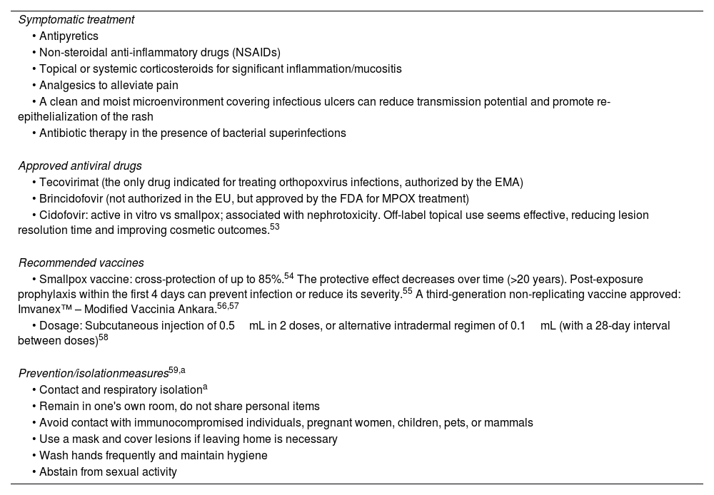
TreatmentAs of today, no specific FDA/EMA-approved treatments exist for patients with MPOX, so symptomatic management remains the cornerstone of therapy (Table 11).
Symptomatic treatment, antiviral drugs, vaccines, and preventive measures recommended for MPOX.
| Symptomatic treatment |
| • Antipyretics |
| • Non-steroidal anti-inflammatory drugs (NSAIDs) |
| • Topical or systemic corticosteroids for significant inflammation/mucositis |
| • Analgesics to alleviate pain |
| • A clean and moist microenvironment covering infectious ulcers can reduce transmission potential and promote re-epithelialization of the rash |
| • Antibiotic therapy in the presence of bacterial superinfections |
| Approved antiviral drugs |
| • Tecovirimat (the only drug indicated for treating orthopoxvirus infections, authorized by the EMA) |
| • Brincidofovir (not authorized in the EU, but approved by the FDA for MPOX treatment) |
| • Cidofovir: active in vitro vs smallpox; associated with nephrotoxicity. Off-label topical use seems effective, reducing lesion resolution time and improving cosmetic outcomes.53 |
| Recommended vaccines |
| • Smallpox vaccine: cross-protection of up to 85%.54 The protective effect decreases over time (>20 years). Post-exposure prophylaxis within the first 4 days can prevent infection or reduce its severity.55 A third-generation non-replicating vaccine approved: Imvanex™ – Modified Vaccinia Ankara.56,57 |
| • Dosage: Subcutaneous injection of 0.5mL in 2 doses, or alternative intradermal regimen of 0.1mL (with a 28-day interval between doses)58 |
| Prevention/isolationmeasures59,a |
| • Contact and respiratory isolationa |
| • Remain in one's own room, do not share personal items |
| • Avoid contact with immunocompromised individuals, pregnant women, children, pets, or mammals |
| • Use a mask and cover lesions if leaving home is necessary |
| • Wash hands frequently and maintain hygiene |
| • Abstain from sexual activity |
EMA: European Medicines Agency; FDA: Food and Drug Administration; EU: European Union.
This is a rare genital infection caused by the aerobic Gram-negative intracellular bacterium Klebsiella granulomatis. Currently, main foci are found in Papua New Guinea, South Africa, parts of India, and Brazil.60,18
After an incubation period of about 50 days, it is characterized by one or more firm papular lesions that initially grow in size and number later progressing to painless ulcers, predominantly in the genital/perianal region (90%). Donovanosis usually occurs without lymphadenopathy and may occasionally be associated with subcutaneous granulomas (pseudobuboes).61 Lesions are highly vascularized and tend to bleed easily, thus increasing the risk of HIV coinfection.62
Clinical presentationThe most common clinical forms are ulcerative-granulomatous, followed by hypertrophic/verrucous, necrotic, and sclerotic. Extragenital infection (6%) can occur in the pelvic area, intra-abdominal organs, bones, or the oral cavity.61
DiagnosisDiagnosis can be established by visualizing the bacterium using dark-field microscopy or direct microscopy with Giemsa/Leishman/Wright staining (large mononuclear cells with intracellular cysts filled with Donovan bodies), by biopsizing lesion samples, or detecting the DNA through PCR. It is a bacterium that is difficult to culture.61
Contact tracing should cover from 60 days60 to 6 months18,61 prior to symptom onset.
The main differential diagnosis is penile squamous cell carcinoma, which can mimic or complicate the infection. Therefore, biopsy of persistent lesions is recommended.64
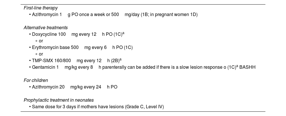
TreatmentThe goal is to halt the progression of lesions; healing typically begins at the ulcer margins, requiring prolonged therapy to allow granulation and re-epithelialization. The minimum treatment duration is 3 weeks, and it should continue until lesions resolve61 (Table 12).
Treatment of granuloma inguinale/donovanosis.
| First-line therapy |
| • Azithromycin 1g PO once a week or 500mg/day (1B; in pregnant women 1D) |
| Alternative treatments |
| • Doxycycline 100mg every 12h PO (1C)a |
| ∘ or |
| • Erythromycin base 500mg every 6h PO (1C) |
| ∘ or |
| • TMP-SMX 160/800mg every 12h (2B)a |
| • Gentamicin 1mg/kg every 8h parenterally can be added if there is a slow lesion response o (1C)a BASHH |
| For children |
| • Azithromycin 20mg/kg every 24h PO |
| Prophylactic treatment in neonates |
| • Same dose for 3 days if mothers have lesions (Grade C, Level IV) |
BASHH: British Association for Sexual Health and HIV; VO: oral route.
Minimum duration: 3 weeks and until complete healing of lesions.
In this chapter, we have outlined the main epidemiological, clinical, and therapeutic characteristics of ulcerative STIs in a practical and straightforward manner for easy access by health care professionals dedicated to the management of STIs. Proper diagnosis will facilitate early treatment, ultimately leading to a reduction in transmission rates.
FundingNone declared.


